

Can the winter have an impact on one’s eyes?
As the days get shorter, can there be an effect on ocular health? In this article, Gurjeet Jutley looks at the typical eye problems associated with the winter months and offers hints and tips on how to cope.
Winter eye problems
A range of specific issues can create problems for our eyes during the winter months. These include:
● Dry eyes – central heating and being indoors can lead to our eyes becoming dry and itchy.
● Teary eyes – cold winds and low temperatures can all irritate our eyes and make us overproduce tears.
● Digital eye strain – as we spend more time indoors we can spend even more time looking at screens.
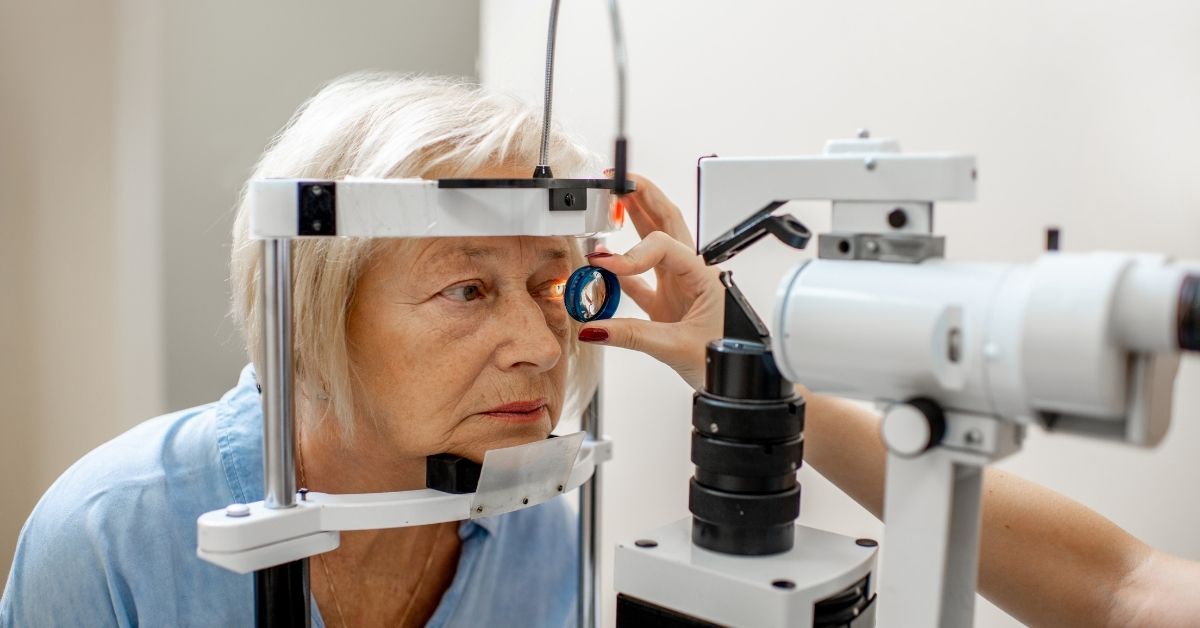
● Contact lenses in the winter – Harsh winds and cold temperature and with the lack of moisture in the air, wearing contact lenses can become uncomfortable and lead to irritation.
Ways to look after your eyes during the winter
As always, general health endeavours reap multiple rewards for all the tissues of the body.
For example:
Drink plenty of water
During the colder months, many of us drink more hot drinks. These are not always hydrating and, in some cases, can dehydrate us further, particularly in the case of caffeinated drinks. Drinking plenty of fresh water helps to keep our eyes, skin and the rest of our body adequately hydrated. This is as essential to our body’s vital functions through the winter as much as it is in the summer.
Look after your diet
We are all human and during the festive months we reach out for carbohydrate and sugar-heavy foods! While there’s nothing wrong with this in moderation it’s important to ensure you eat plenty of fresh fruit and vegetables.
Various vitamins are beneficial in maintaining good eye health:
● Vitamin A protects the cornea and it may lower the risk of cataracts and macular degeneration. Found in cheese, eggs, oily fish and yoghurts.
● Vitamin E is an important antioxidant, that helps to protect your body’s cells, including those in your eyes. Soya, corn and olive oil are great sources of Vitamin E.
● Vitamin C is required to make collagen a protein that provides structure to your eyes. Oranges, broccoli and red and green peppers can help you get enough.
As well as vitamins, Omega-3 fatty acids help to form the cells of the eyes and also contain anti-inflammatory properties.
Use a dehumidifier
Heaters and radiators can make the air in your home dry which can lead to eye irritation. To help keep your eyes moist it can be helpful to use a dehumidifier in the home. Also, reducing the radiator temperature can be helpful.
Use eye drops
Lubricating eye drops can help to protect your eyes generally. They can help reduce the drying impact of wind and cold air but are also helpful if you’re spending time in a centrally heated home or workplace. If you’re a contact lens wearer make sure that they’re suitable for lenses, particular emphasis to preservative free.
Wear your glasses
If you’re a glasses wearer the good news is that they offer excellent protection for your eyes against the impact of winter cold and drying winds. Those of us who enjoy skiing, sunglasses can give UV protection and avoid photokeratitis.


Recent Comments